We use cookies to enhance the usability of our website. If you continue, we'll assume that you are happy to receive all cookies. More information. Don't show this again.
General description of the gene and the encoded protein(s) using information from HGNC and Ensembl, as well as predictions made by the Human Protein Atlas project.
Gene namei
Official gene symbol, which is typically a short form of the gene name, according to HGNC.
All transcripts of all genes have been analyzed regarding the location(s) of corresponding protein based on prediction methods for signal peptides and transmembrane regions.
Genes with at least one transcript predicted to encode a secreted protein, according to prediction methods or to UniProt location data, have been further annotated and classified with the aim to determine if the corresponding protein(s) are secreted or actually retained in intracellular locations or membrane-attached.
Remaining genes, with no transcript predicted to encode a secreted protein, will be assigned the prediction-based location(s).
The annotated location overrules the predicted location, so that a gene encoding a predicted secreted protein that has been annotated as intracellular will have intracellular as the final location.
Gene information from Ensembl and Entrez, as well as links to available gene identifiers are displayed here. Information was retrieved from Ensembl if not indicated otherwise.
Chromosome
7
Cytoband
q33
Chromosome location (bp)
137227341 - 137343774
Number of transcriptsi
Number of protein-coding transcripts from the gene as defined by Ensembl.
The Structure section provides predicted structures from the Alphafold protein structure database and available experimental structures from Protein Data Bank (PDB).
In the Structure drop-down menu all experimental structures from PDB are available for selection and display. The structures are displayed using the NGL Viewer and can be zoomed-in and rotated either manually or by checking the Autorotate box. The Color scheme can be selected to show the residue index, chain name or confidence score (as B-factors and pLDDT score for experimental and predicted structures, respectively). The positions for available antigen sequences in the structure are shown if Antigens is turned to ON, and the Variants slider can be used to show the positions of clinical and population variants.https://github.com/nglviewer/ngl
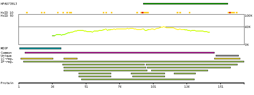
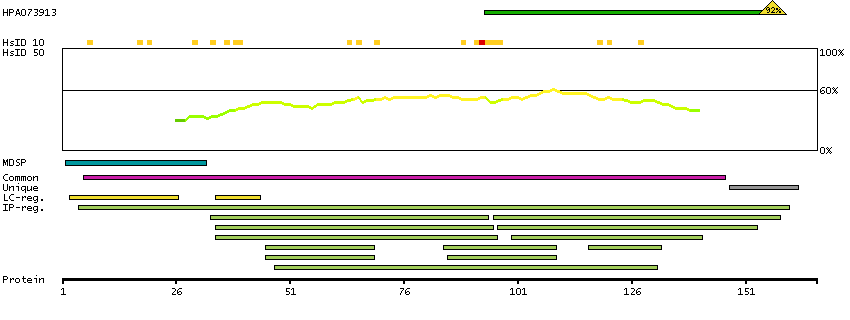
The protein browser displays the antigen location on the target protein(s) and the features of the target protein. The tabs at the top of the protein view section can be used to switch between the different splice variants to which an antigen has been mapped.
At the top of the view, the position of the antigen (identified by the corresponding HPA identifier) is shown as a green bar. A yellow triangle on the bar indicates a <100% sequence identity to the protein target.
Below the antigens, the maximum percent sequence identity of the protein to all other proteins from other human genes is displayed, using a sliding window of 10 aa residues (HsID 10) or 50 aa residues (HsID 50). The region with the lowest possible identity is always selected for antigen design, with a maximum identity of 60% allowed for designing a single-target antigen (read more).
The curve in blue displays the predicted antigenicity i.e. the tendency for different regions of the protein to generate an immune response, with peak regions being predicted to be more antigenic.The curve shows average values based on a sliding window approach using an in-house propensity scale. (read more).
If a signal peptide is predicted by a majority of the signal peptide predictors SPOCTOPUS, SignalP 4.0, and Phobius (turquoise) and/or transmembrane regions (orange) are predicted by MDM, these are displayed.
Low complexity regions are shown in yellow and InterPro regions in green. Common (purple) and unique (grey) regions between different splice variants of the gene are also displayed (read more), and at the bottom of the protein view is the protein scale.
PTN-201
PTN-202
PROTEIN INFORMATIONi
The protein information section displays alternative protein-coding transcripts (splice variants) encoded by this gene according to the Ensembl database.
The ENSP identifier links to the Ensembl website protein summary, while the ENST identifier links to the Ensembl website transcript summary for the selected splice variant. The data in the UniProt column can be expanded to show links to all matching UniProt identifiers for this protein.
The protein classes assigned to this protein are shown if expanding the data in the protein class column. Parent protein classes are in bold font and subclasses are listed under the parent class.
The Gene Ontology terms assigned to this protein are listed if expanding the Gene ontology column. The length of the protein (amino acid residues according to Ensembl), molecular mass (kDalton), predicted signal peptide (according to a majority of the signal peptide predictors SPOCTOPUS, SignalP 4.0, and Phobius) and the number of predicted transmembrane region(s) (according to MDM) are also reported.
MEMSAT3 predicted membrane proteins TMHMM predicted membrane proteins Predicted secreted proteins Secreted proteins predicted by MDSEC SignalP predicted secreted proteins Phobius predicted secreted proteins SPOCTOPUS predicted secreted proteins Cancer-related genes Candidate cancer biomarkers Mapped to neXtProt neXtProt - Evidence at protein level Protein evidence (Kim et al 2014) Protein evidence (Ezkurdia et al 2014)
Show all
GO:0001503 [ossification] GO:0001889 [liver development] GO:0002232 [leukocyte chemotaxis involved in inflammatory response] GO:0002690 [positive regulation of leukocyte chemotaxis] GO:0004864 [protein phosphatase inhibitor activity] GO:0005178 [integrin binding] GO:0005515 [protein binding] GO:0005539 [glycosaminoglycan binding] GO:0005576 [extracellular region] GO:0005604 [basement membrane] GO:0005615 [extracellular space] GO:0005737 [cytoplasm] GO:0005783 [endoplasmic reticulum] GO:0005886 [plasma membrane] GO:0007165 [signal transduction] GO:0007185 [transmembrane receptor protein tyrosine phosphatase signaling pathway] GO:0007229 [integrin-mediated signaling pathway] GO:0007399 [nervous system development] GO:0007406 [negative regulation of neuroblast proliferation] GO:0007420 [brain development] GO:0007507 [heart development] GO:0007612 [learning] GO:0007613 [memory] GO:0008083 [growth factor activity] GO:0008201 [heparin binding] GO:0008284 [positive regulation of cell population proliferation] GO:0008360 [regulation of cell shape] GO:0009986 [cell surface] GO:0010594 [regulation of endothelial cell migration] GO:0010811 [positive regulation of cell-substrate adhesion] GO:0010976 [positive regulation of neuron projection development] GO:0010996 [response to auditory stimulus] GO:0014823 [response to activity] GO:0016020 [membrane] GO:0016525 [negative regulation of angiogenesis] GO:0019901 [protein kinase binding] GO:0021510 [spinal cord development] GO:0021549 [cerebellum development] GO:0021794 [thalamus development] GO:0030282 [bone mineralization] GO:0030324 [lung development] GO:0030336 [negative regulation of cell migration] GO:0030501 [positive regulation of bone mineralization] GO:0030902 [hindbrain development] GO:0031104 [dendrite regeneration] GO:0031594 [neuromuscular junction] GO:0031641 [regulation of myelination] GO:0032355 [response to estradiol] GO:0032515 [negative regulation of phosphoprotein phosphatase activity] GO:0032570 [response to progesterone] GO:0032991 [protein-containing complex] GO:0034644 [cellular response to UV] GO:0035373 [chondroitin sulfate proteoglycan binding] GO:0035374 [chondroitin sulfate binding] GO:0036120 [cellular response to platelet-derived growth factor stimulus] GO:0038085 [vascular endothelial growth factor binding] GO:0042246 [tissue regeneration] GO:0042493 [response to drug] GO:0043065 [positive regulation of apoptotic process] GO:0043113 [receptor clustering] GO:0043394 [proteoglycan binding] GO:0043932 [ossification involved in bone remodeling] GO:0044849 [estrous cycle] GO:0045446 [endothelial cell differentiation] GO:0045545 [syndecan binding] GO:0045597 [positive regulation of cell differentiation] GO:0045778 [positive regulation of ossification] GO:0045837 [negative regulation of membrane potential] GO:0046697 [decidualization] GO:0048167 [regulation of synaptic plasticity] GO:0048471 [perinuclear region of cytoplasm] GO:0048477 [oogenesis] GO:0048680 [positive regulation of axon regeneration] GO:0048714 [positive regulation of oligodendrocyte differentiation] GO:0050680 [negative regulation of epithelial cell proliferation] GO:0051781 [positive regulation of cell division] GO:0060041 [retina development in camera-type eye] GO:0060221 [retinal rod cell differentiation] GO:0060253 [negative regulation of glial cell proliferation] GO:0060291 [long-term synaptic potentiation] GO:0071305 [cellular response to vitamin D] GO:0071407 [cellular response to organic cyclic compound] GO:0071456 [cellular response to hypoxia] GO:0072201 [negative regulation of mesenchymal cell proliferation] GO:0098793 [presynapse] GO:0098794 [postsynapse] GO:0140059 [dendrite arborization] GO:1900006 [positive regulation of dendrite development] GO:1900272 [negative regulation of long-term synaptic potentiation] GO:1903706 [regulation of hemopoiesis] GO:1904373 [response to kainic acid] GO:1904389 [rod bipolar cell differentiation] GO:1904391 [response to ciliary neurotrophic factor] GO:1904395 [positive regulation of skeletal muscle acetylcholine-gated channel clustering] GO:1904397 [negative regulation of neuromuscular junction development] GO:1904399 [heparan sulfate binding] GO:1990089 [response to nerve growth factor] GO:2000036 [regulation of stem cell population maintenance] GO:2000347 [positive regulation of hepatocyte proliferation] GO:2000738 [positive regulation of stem cell differentiation]
 The Human Protein Atlas project is funded