We use cookies to enhance the usability of our website. If you continue, we'll assume that you are happy to receive all cookies. More information. Don't show this again.
VIP
SECTIONS
  • TISSUE
  • BRAIN
  • SINGLE CELL TYPE
  • TISSUE CELL TYPE
  • PATHOLOGY
  • DISEASE
  • IMMUNE CELL
  • BLOOD PROTEIN
  • SUBCELLULAR
  • CELL LINE
  • STRUCTURE
  • METABOLIC
ABOUT
  • INTRODUCTION
  • HISTORY
  • ORGANIZATION
  • PUBLICATIONS
  • ANTIBODY SUBMISSION
  • ANTIBODY AVAILABILITY
  • ACKNOWLEDGMENTS
  • CONTACT
NEWS
  • NEWS ARTICLES
  • PRESS ROOM
LEARN
  • DICTIONARY
  • PROTEIN CLASSES
  • PROTEIN EVIDENCE
  • METHODS
  • EDUCATIONAL VIDEOS
DATA
  • DOWNLOADABLE DATA
  • PUBLICATION DATA
  • RELEASE HISTORY
  • SARS-COV-2
HELP
  • ANTIBODY VALIDATION
  • ASSAYS & ANNOTATION
  • DISCLAIMER
  • HELP & FAQ
  • PRIVACY STATEMENT
  • LICENCE & CITATION
Fields »
Search result

Field
Term
Gene name
Class
Subclass
Class
Keyword
Chromosome
External id
Tissue
Cell type
Expression
Patient ID
Tissue
Category
Cluster
Reliability
Brain region
Category
Brain region
Category
Brain region
Category
Reliability
Cell type
Category
Cluster
Tissue
Cell type
Enrichment
Cancer
Prognosis
Cancer
Category
Cell type
Category
Cell lineage
Category
Cluster
Annotation
Disease
Location
Searches
Location
Cell line
Type
Phase
Reliability
Cancer type
Category
Cluster
Pathway
Category
Score
Score
Score
Validation
Validation
Validation
Validation
Antibodies
Protein structure
In atlas
Column


  • SUMMARY

  • TISSUE

  • BRAIN

  • SINGLE CELL

  • TISSUE CELL

  • PATHOLOGY

  • DISEASE

  • IMMUNE

  • BLOOD

  • SUBCELL

  • CELL LINE

  • STRUCTURE

  • METABOLIC

  • VIP
BRAIN TISSUES Show tissue menu
AMYGDALA BASAL GANGLIA CEREBELLUM CEREBRAL CORTEX HIPPOCAMPAL FORMATION HYPOTHALAMUS MEDULLA OBLONGATA MIDBRAIN
OLFACTORY BULB PITUITARY GLAND PONS RETINA SPINAL CORD THALAMUS WHITE MATTER
PRIMARY MOUSE DATA
ANTIBODIES
AND
VALIDATION
Brain proteome
MOUSE BRAIN TISSUE
Gene description Vasoactive intestinal peptide
Mouse gene ENSMUSG00000019772 (version 10)
Annotation summary 
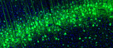
Vasoactive Intestinal Peptide (VIP) is a peptide hormone produced mainly in the gut, pancreas and brain where it activates gene expression pathways involved in circadian rhythm. VIP RNA shows higher expression in the appendix, colon and small intestine among all human tissues, with levels in the nervous system being comparatively lower. In the mouse brain, RNA levels are widespread across many forebrain regions, while, on the protein level, moderate to strong intensity immunostaining of dense axonal networks is noticed in multiple but distinct brain regions. Strong intensity dense axonal staining is observed in the laterodorsal and mediodorsal BNST nuclei, in the suprachiasmatic nucleus, periventricularly in the anterior hypothalamus, including the paraventricular nucleus, and in the central amygdala. Moderate-intensity axonal staining is observed in the septum, hippocampus, neocortical areas and in the piriform cortex. In addition, moderate-intensity immunostaining of neuronal cell bodies is noticed in subsets of neurons throughout the neocortex and hippocampus, in the periaqueductal gray matter as well as in the olfactory bulb external plexiform layer.
Positive cells and structuresi

Manually selected location of the protein positivity, observed by immunofluorescence staining in mouse brain.

Read more
Soma and axon in neurons.
FRONT BACK
Olfactory bulb
Cerebral cortex
Basal ganglia
Protein
Relative fluorescence intensityi

Protein expression in mouse brain using immunofluorescence is reported as mean fluorescence intensity .

Read more
Mouse brain region
255
0
Cerebral cortex
Frontal association cortex (fra)
Motor cortex (m)
Cingulate cortex (cg)
Piriform cortex, L1 (pirl1)
Piriform cortex, L2 (pirl2)
Piriform cortex, L3 (pirl3)
Insular cortex (i)
Somatosensory cortex (s)
Retrosplenial granular cortex (rsg)
Parietal association cortex (p) N/A
Entorhinal cortex (ent)
Visual cortex (v)
Olfactory bulb
Anterior olfactory nucleus (aon)
Granule cell layer (gro)
Internal plexiform layer (ipl)
Mitral cell layer (mi)
Glomerular layer (gl)
Rostral migratory stream (rms)
External plexiform layer (epl)
External plexiform layer of the accessory OB (epla)
Granule cell layer of the accessory OB (gra)
Glomerular layer of the accessory OB (gla)
Hippocampus
Polymorph layer of the dentate gyrus (podg)
Molecular layer of the dentate gyrus (modg)
Granular dentate gyrus (grdg)
CA1 - oriens layer (ca1or)
CA1 - pyramidal layer (ca1py)
CA1 - radiatum layer (ca1ra)
CA2 - oriens layer (ca2or)
CA2 - pyramidal layer (ca2py)
CA2 - radiatum layer (ca2ra)
CA3 - oriens layer (ca3or)
CA3 - pyramidal layer (ca3py)
CA3 - radiatum layer (ca3ra)
Stratum lucidum (slu)
Lacunosum moleculare (lmol)
Subiculum (sub)
Amygdala
Nucleus of the lateral olfactory tract (lot) N/A
Basal medial amygdaloid nucleus (bma)
Basal lateral amygdala* (*bla) N/A
Basal lateral amygdaloid nucleus (bla)
Cortical amygdala (aco)
Central amygdala (ce)
Medial amygdaloid nucleus (mea)
Thalamus
Medial geniculate nucleus (mg)
Parafascicular thalamic nucleus (pf) N/A
Pregeniculate nucleus (pg)
Stria terminalis (st)
Zona incerta (zi)
Anterodorsal thalamic nucleus (ad)
Reticular thalamic nucleus (rt)
Vental anterior thalamic nucleus (va) N/A
Medial habenular nucleus (mhb)
Laterodorsal thalamic area (ld)
Paraventricular thalamic nucleus (pv)
Central medial thalamic area (cm)
Ventral lateral thalamic area (vl) N/A
Ventral medial thalamic area (vm)
Lateral habenulal nucleus (lhb)
Ventral posterior thalamus (vpt)
Anterior pretactal nucleus (apt) N/A
Retromammillary nucleus (rm)
Hypothalamus
Dorsal tuberomammillary nucleus (dtm) N/A
Mammillary nucleus (mn)
Periventricular hypothalamic nucleus (pe)
Supraoptic nucleus (so)
Tuberal nucleus (tu)
Ventral tuberomammillary nucleus (vtm)
Lateral preoptic area (lpo)
Medial preoptic area (mpo)
Suprachiasmatic nucleus (sch)
Paraventricular hypothalamic nucleus (pa)
Anterior hypothalamic area, central (ahc)
Ventral medial hypothalamic nucleus (vmh)
Ventral medial hypothalamic nucleus* (*vmh) N/A
Arcuate nucleus (arc)
Arcuate nucleus* (*arc) N/A
Peduncular part of lateral hypothalmus (plh)
Dorsal medial hypothalamic nucleus (dm)
Stria terminalis* (*st) N/A
Cerebellum
Vestibulocerebellar nucleus (vecb) N/A
Moleuclar layer of the cerebellum (cemol)
Purkinje layer of the cerebellum (cepur)
Granular layer of the cerebellum (cegr)
Pons
Barrington's nucleus (bar) N/A
Koelliker-fuse nucleus (kf) N/A
Motor tregiminal nucleus (5n)
Parabrachial nucleus (pbp)
Principle sensory trigeminal nucleus (pr5)
Locus coeruleus (lc)
Pontine nucleus (pn)
Vestibular nucleus (ve)
Pontine reticular nucleus, oral (pno)
Lateral lemniscus (ll)
Superior paraolivary nucleus (spo)
Circumventricular organs
Subcommissural organ (sco)
Subfornical organ (sfo)
Median eminence (me) N/A
Medulla (ap)

Antibodies in assayi

The internal antibody ID for the antibody/antibodies available for this specific target, click link for more antibody information.

CAB079780

Contact

  • NEWS ARTICLES
  • PRESS ROOM
  • contact@proteinatlas.org

The Project

  • INTRODUCTION
  • ORGANIZATION
  • PUBLICATIONS

The Human Protein Atlas

  • DOWNLOADABLE DATA
  • LICENCE & CITATION
  • HELP & FAQ
The Human Protein Atlas project is funded
by the Knut & Alice Wallenberg Foundation.