We use cookies to enhance the usability of our website. If you continue, we'll assume that you are happy to receive all cookies. More information. Don't show this again.
CHCHD7
SECTIONS
  • TISSUE
  • BRAIN
  • SINGLE CELL TYPE
  • TISSUE CELL TYPE
  • PATHOLOGY
  • DISEASE
  • IMMUNE CELL
  • BLOOD PROTEIN
  • SUBCELLULAR
  • CELL LINE
  • STRUCTURE
  • METABOLIC
ABOUT
  • INTRODUCTION
  • HISTORY
  • ORGANIZATION
  • PUBLICATIONS
  • ANTIBODY SUBMISSION
  • ANTIBODY AVAILABILITY
  • ACKNOWLEDGMENTS
  • CONTACT
NEWS
  • NEWS ARTICLES
  • PRESS ROOM
LEARN
  • DICTIONARY
  • PROTEIN CLASSES
  • PROTEIN EVIDENCE
  • METHODS
  • EDUCATIONAL VIDEOS
DATA
  • DOWNLOADABLE DATA
  • PUBLICATION DATA
  • RELEASE HISTORY
  • SARS-COV-2
HELP
  • ANTIBODY VALIDATION
  • ASSAYS & ANNOTATION
  • DISCLAIMER
  • HELP & FAQ
  • PRIVACY STATEMENT
  • LICENCE & CITATION
Fields ยป
Search result

 
Field
All
Term
Gene name
Class
Blood group antigen proteins
Subclass
 
Class
 
Keyword
Chromosome
External id
Tissue
 
Cell type
 
Expression
Patient ID
Tissue
Category
Cluster
Reliability
Brain region
Category
Brain region
Category
Brain region
Category
Reliability
Cell type
Category
Cluster
Tissue
 
Cell type
Enrichment
Cancer
Prognosis
Cancer
Category
Cell type
Category
Cell lineage
Category
Cluster
Annotation
Disease
Location
Searches
Location
Cell line
Type
Phase
Reliability
Cancer type
Category
Cluster
Pathway
Category
Score
Score
Score
Validation
Validation
Validation
Validation
Antibodies
Yes
Protein structure
In atlas
Column
Gene


                        • SPECIFICITY
                        • DISTRIBUTION
                        • EXPRESSION
                        • SPECIFICITY
                        • DISTRIBUTION
                        • EXPRESSION
                        • SPECIFICITY
                        • DISTRIBUTION
                        • EXPRESSION
                        • SPECIFICITY
                        • DISTRIBUTION
                        • EXPRESSION
                        • SPECIFICITY
                        • DISTRIBUTION
                        • EXPRESSION
                          • SPECIFICITY
                          • DISTRIBUTION
                          • EXPRESSION
                          • SPECIFICITY
                          • DISTRIBUTION
                          • EXPRESSION
                          • SPECIFICITY
                          • DISTRIBUTION
                          • EXPRESSION
                          • RELIABILITY
                          • SUBCELLULAR VARIATION
                          • MULTILOCALIZATION
                          • OTHER
                          • SPECIFICITY
                          • DISTRIBUTION
                          • EXPRESSION
                          • Check all
                          • Uncheck all
                                • SUMMARY

                                • TISSUE

                                • BRAIN

                                • SINGLE CELL

                                • TISSUE CELL

                                • PATHOLOGY

                                • DISEASE

                                • IMMUNE

                                • BLOOD

                                • SUBCELL

                                • CELL LINE

                                • STRUCTURE

                                • METABOLIC

                                • CHCHD7
                                TISSUE PRIMARY DATA TISSUES Show tissue menu
                                ADIPOSE TISSUE ADRENAL GLAND AMYGDALA APPENDIX BASAL GANGLIA BONE MARROW BREAST BRONCHUS CEREBELLUM CEREBRAL CORTEX CERVIX CHOROID PLEXUS COLON DUODENUM ENDOMETRIUM
                                EPIDIDYMIS ESOPHAGUS FALLOPIAN TUBE GALLBLADDER HEART MUSCLE HIPPOCAMPAL FORMATION HYPOTHALAMUS KIDNEY LIVER LUNG LYMPH NODE MEDULLA OBLONGATA MIDBRAIN ORAL MUCOSA OVARY
                                PANCREAS PARATHYROID GLAND PITUITARY GLAND PLACENTA PONS PROSTATE RECTUM RETINA SALIVARY GLAND SEMINAL VESICLE SKELETAL MUSCLE SKIN SMALL INTESTINE SMOOTH MUSCLE SOFT TISSUE
                                SPINAL CORD SPLEEN STOMACH TESTIS THALAMUS THYMUS THYROID GLAND TONGUE TONSIL URINARY BLADDER VAGINA WHITE MATTER
                                ANTIBODIES
                                AND
                                VALIDATION
                                Dictionary
                                Tissue proteome
                                ANTIBODY PRIMARY DATAi

                                The primary data page shows the detailed antibody staining in 76 different cell types for each analyzed antibody. In addition, for each cell type, a knowledge-based annotated protein expression score based on all analyzed antibodies is displayed furthest to the right. The images and annotations can be accessed by clicking on the tissue name or score bar. More information can be found in Assays & Annotation.

                                The tissues and cell types can be ordered by organ, cell type or alphabetically. If ordered by organ, the tissues are presented in groups according to functional features. If ordered by cell type, the cells are presented in groups related to origin.

                                For histological reference, visit the histological dictionary.

                                Read more
                                Antibodies in assayi

                                The internal antibody ID for the antibody/antibodies available for this specific target, click link for more antibody information.

                                HPA050783
                                Tissue presentation order  
                                Organ
                                Cell type
                                Alphabetical
                                Tissue Antibody stainingi

                                This score describes the level of antibody staining observed in the annotated cell types as not detected (n), low (l), medium (m) or high (h). It is based on the staining intensity and fraction of stained cells. The images and annotations can be accessed by clicking on the tissue name or score bar. More information can be found in Assays & Annotation.

                                Read more
                                Annotated protein expressioni

                                This score describes a knowledge-based best estimate of the protein expression in the annotated cell types as not detected (n), low (l), medium (m) or high (h). It is a result of stringent evaluation of immunohistochemical staining pattern, RNA data from internal and external sources and available protein/gene characterization data. More information can be found in Assays & Annotation.

                                Read more

                                HPA050783

                                h
                                m
                                l
                                n
                                h
                                m
                                l
                                n
                                Brain
                                Cerebral cortex Neuronal cells
                                Cerebral cortex Endothelial cells
                                Cerebral cortex Glial cells
                                Cerebral cortex Neuropil
                                Hippocampus Neuronal cells
                                Hippocampus Glial cells
                                Caudate Neuronal cells
                                Caudate Glial cells
                                Cerebellum Purkinje cells
                                Cerebellum Cells in granular layer
                                Cerebellum Cells in molecular layer
                                Endocrine tissues
                                Thyroid gland Glandular cells
                                Parathyroid gland Glandular cells
                                Adrenal gland Glandular cells
                                Respiratory system
                                Nasopharynx Respiratory epithelial cells
                                N/A
                                Bronchus Basal cells
                                Bronchus Ciliated cells (cell body)
                                Bronchus Ciliated cells (cilia axoneme)
                                Bronchus Ciliated cells (ciliary rootlets)
                                Bronchus Ciliated cells (tip of cilia)
                                Bronchus Goblet cells
                                Lung Alveolar cells
                                Lung Macrophages
                                Proximal digestive tract
                                Oral mucosa Squamous epithelial cells
                                Salivary gland Glandular cells
                                Esophagus Squamous epithelial cells
                                Gastrointestinal tract
                                Stomach 1 Glandular cells
                                Stomach 2 Glandular cells
                                Duodenum Glandular cells
                                Small intestine Glandular cells
                                Colon Endothelial cells
                                Colon Glandular cells
                                Colon Peripheral nerve/ganglion
                                Rectum Glandular cells
                                Liver & Gallbladder
                                Liver Hepatocytes
                                Liver Cholangiocytes
                                Gallbladder Glandular cells
                                Pancreas
                                Pancreas Exocrine glandular cells
                                Pancreas Pancreatic endocrine cells
                                Kidney & Urinary bladder
                                Kidney Cells in glomeruli
                                Kidney Cells in tubules
                                Urinary bladder Urothelial cells
                                Male reproductive system
                                Testis Cells in seminiferous ducts
                                Testis Leydig cells
                                Epididymis Glandular cells
                                Prostate Glandular cells
                                Seminal vesicle Glandular cells
                                Breast and female reproductive system
                                Breast Adipocytes
                                Breast Glandular cells
                                Breast Myoepithelial cells
                                Vagina Squamous epithelial cells
                                Cervix Squamous epithelial cells
                                N/A
                                Cervix Glandular cells
                                Endometrium 1 Glandular cells
                                Endometrium 1 Cells in endometrial stroma
                                Endometrium 2 Glandular cells
                                Endometrium 2 Cells in endometrial stroma
                                Fallopian tube Ciliated cells (cell body)
                                Fallopian tube Ciliated cells (cilia axoneme)
                                Fallopian tube Ciliated cells (ciliary rootlets)
                                Fallopian tube Ciliated cells (tip of cilia)
                                Fallopian tube Non-ciliated cells
                                Ovary Follicle cells
                                N/A
                                Ovary Ovarian stroma cells
                                Placenta Trophoblastic cells
                                Placenta Decidual cells
                                N/A
                                Muscle tissues
                                Heart muscle Cardiomyocytes
                                Skeletal muscle Myocytes
                                Smooth muscle Smooth muscle cells
                                Connective & Soft tissue
                                Adipose tissue Adipocytes
                                Soft tissue 1 Peripheral nerve
                                Soft tissue 1 Chondrocytes
                                N/A
                                Soft tissue 1 Fibroblasts
                                Soft tissue 2 Fibroblasts
                                Soft tissue 2 Chondrocytes
                                N/A
                                Soft tissue 2 Peripheral nerve
                                N/A
                                Skin
                                Skin 1 Fibroblasts
                                Skin 1 Keratinocytes
                                Skin 1 Langerhans
                                Skin 1 Melanocytes
                                Skin 2 Epidermal cells
                                N/A
                                Bone marrow & Lymphoid tissues
                                Appendix Glandular cells
                                Appendix Lymphoid tissue
                                Bone marrow Hematopoietic cells
                                Lymph node Germinal center cells
                                Lymph node Non-germinal center cells
                                Tonsil Germinal center cells
                                Tonsil Non-germinal center cells
                                Tonsil Squamous epithelial cells
                                Spleen Cells in white pulp
                                Spleen Cells in red pulp
                                 
                                Staining summaryi

                                Summarizing texts describe the staining pattern for each antibody based on staining intensity, fraction of stained cells and subcellular localization. More information can be found in Assays & Annotation.

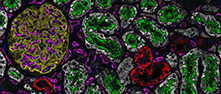
                                Parathyroid gland and a subset of cells in red pulp of spleen displayed strong cytoplasmic positivity. Cardiac myocytes showed moderate cytoplasmic and membranous staining. Gastrointestinal tract, hepatocytes, renal tubules and the neuronal cells showed moderate cytoplasmic staining. Remaining normal tissues were negative.

                                Contact

                                • NEWS ARTICLES
                                • PRESS ROOM
                                • contact@proteinatlas.org

                                The Project

                                • INTRODUCTION
                                • ORGANIZATION
                                • PUBLICATIONS

                                The Human Protein Atlas

                                • DOWNLOADABLE DATA
                                • LICENCE & CITATION
                                • HELP & FAQ
                                The Human Protein Atlas project is funded
                                by the Knut & Alice Wallenberg Foundation.